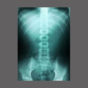
Spinal stenosis x-ray is a preliminary diagnostic test which might show definitive evidence of certain types of central canal and foraminal narrowing. X-rays are popular due to their easy and inexpensive nature, but are extremely limited in what they can visualize. In the case of diagnosing spinal stenosis, x-rays are not typically the best bet, compared to other more advanced forms of diagnostic imaging, such as MRI. However, these diagnostic imaging devices can still be useful to patients in particular circumstances.
This discussion will detail the usefulness and limitations of x-ray technology in association with spinal stenosis conditions.
Spinal Stenosis X-Ray Diagnostic Limitations
X-rays only accurately visualize bones. They can not detail soft tissues and are therefore very limited in what they can diagnose in the spine. Most significant arthritic changes, such as osteophytes, will show up on x-rays. However, soft tissue conditions, such as herniated discs and ligamentum flavum hypertrophy will not be detailed at all.
Additionally, even the bone issues which show up will not be very useful, since the soft neurological tissues which may or may not be affected will not appear on the images. These include the actual spinal cord and the spinal nerves.
X-Ray Diagnostic Usefulness
X-rays are most useful for diagnosing suspected serious bone issues, such as fractures, spondylolisthesis or abnormal curvatures. X-rays are usually the first line of diagnostic testing performed after a trauma, such as a fall or car accident.
X-rays are particularly ineffective at diagnosing most sources of dorsalgia and are even used as convincing evidence that treatment is needed, even when the patient does not actually understand the image presented to them. We see this often with chiropractors who take x-rays in their office. They inevitably present pictures of a lumber spine or cervical spine with narrowed disc spaces and make a big deal of these images.
Of course, degenerative disc disease is normal and universal in these areas, unbeknownst to many patients. The purpose of this practice is to enlist the patient into long-term and profitable treatment, for a condition which is highly unlikely to be the source of any pain. For shame.
Spinal Stenosis X-Ray Considerations
Unless you or your doctor suspects a fracture or spinal curvature as the source of your pain, then skipping an x-ray altogether may not be a bad idea. They are simply not worth the time or effort for many patients.
Once thought to be a miracle of technology, x-rays are now a dinosaur in the diagnostic sector of medicine and should be replaced universally by CT, MRI and new forms of computer assisted 3 dimensional imaging in the near future. Until that day, they are still here, but usually have little meaning to patients with chronic back or neck ache.
For spinal stenosis patients who are suffering from bone-related changes, x-rays be provide some evidence of the causative process for pain, maybe enough to warrant a more detailed MRI scan. For most patients, however, x-rays will not provide much more than an educated guess as to the source of symptoms, if even that.
Spinal Stenosis > Spinal Stenosis Diagnosis > Spinal Stenosis X-Ray


