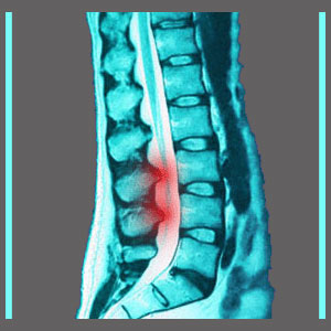
Spinal canal stenosis is another commonly applied diagnostic term used to signify degenerative narrowing changes in the central canal. This completely normal condition generally occurs in the lumbar or cervical areas of the spine, but can also reside in the thoracic region in much rarer instances. The term stenosis means narrowing and the term spinal canal describes the open passageway through which the anatomical mass of soft tissues and nerves which runs through the vertebral bones, carrying blood, nerve signals and cerebral spinal fluid. The primary component contained within the central spinal canal is surely the spinal cord.
This article will provide an overview of central narrowing of the interior canal space and will help patients to further research their specific diagnosis.
Spinal Canal Stenosis Description
The central canal is easy to imagine. Just picture the vertebrae having a hollow tunnel in between the mass of the bone, called the vertebral body, and the fins which project off the back of the vertebrae, called spinous processes. This space is occupied by several types of tissue, including ligaments, protective membranes, nerve tissue, blood vessels and the spinal cord, running from the base of the brain until the upper lumbar region.
In the lumbar area, the spinal cord branches off to form the cauda equina, which is simply a network of individual nerve roots which resembles its namesake, a horse’s tail. This cauda equina slowly decreases in size as nerve rots continue to branch off an exit the canal throughout the lumbar, sacral and coccygeal foramen.
Mechanics of Spinal Stenosis
In a spinal stenosis condition, this central passageway is no longer clear and free from obstructions. In fact, the central canal is instead narrowed to some degree by one or more structural components. The most common causes of spinal stenosis in the central canal include:
Herniated discs can compress the soft tissues within the spinal canal or even threaten the functionality of the spinal cord itself. Usually symptomatic intervertebral bulges are central or paramedial herniations.
Arthritic changes can take place within the spinal canal, on the vertebral bodies or in the facet joints, leading to central stenosis or even neuroforaminal stenosis.
Scoliosis and abnormal front-to-back spinal curvatures, such as hyperlordosis or hyperkyphosis, can enact a stenosis condition in the central canal or in one or more neuroforamen.
Spondylolisthesis or other vertebral misalignment issue can create central or neuroforaminal narrowing.
Understanding Spinal Canal Stenosis
Understanding your diagnosis is the first step in finding effective spinal stenosis treatment. So many patients are led astray during the diagnostic process, simply because they have no idea what their doctor is talking about. Even though they may not need any treatment, their ignorance of the occurrence of stenosis makes them easy marks for unethical and greedy care providers who take the opportunity to make some easy money. Other patients really need the treatments, but are not sure about the best path for them and wind up making the potentially worst choice possible, such as unnecessary spinal fusion. This can be the costliest mistake of a person’s life and may enact damage which can never be undone.
Be careful and take your time when discussing and considering stenosis treatments. Most of all, get involved and become proactive in your care. If you simply sit back and allow everything to be decided for you, the odds that the worst case scenario will become reality increase drastically.
Spinal Stenosis > What is Spinal Stenosis? > Spinal Canal Stenosis


