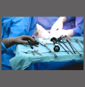
Minimally invasive stenosis treatment may allow for the resolution of stenotic changes in the vertebral canal without incurring excessive damage to healthy tissues. Minimally invasive techniques have become the norm in the surgical treatment of all manner of health issues, but are especially prevalent in the spinal surgery sector. Since stenotic changes exist inside the vertebral canal, deep inside the body, surgical efforts to clear areas of focal stenosis must first access the affected area. Traditionally, this meant utilizing large incisions and muscular dissection to visualize the backbone and then the application of orthopedic surgery techniques to actually cut into the spine and expose the stenotic regions. This type of surgery was barbaric, traumatic, dangerous, risky and typically left large scars. The recuperation time was always long and painful.
Modern surgical practices center on the use of minimally invasive technologies and less traumatic approaches to care that can still address the problematic stenotic regions of the vertebral canal, without upsetting the surrounding anatomy more than absolutely necessary. The goals of minimally invasive therapy include decreased healing time, minimized blood loss, less risk, lower rate of complications and improved patient comfort throughout the entire care process.
The dissertation examines the latest surgical procedures for the treatment of spinal stenosis using the least invasive means of treatment possible. We will detail the techniques used to resolve both central canal and neuroforaminal stenosis.
Minimally Invasive Stenosis Treatment Types
Technological advancements are the main factors that make minimally invasive procedures possible in the modern surgical arena. There are many different ways in which technology can be utilized to reduce trauma during operative interventions, including any and all of the following factors that are commonly involved in minimally invasive stenosis operations:
Fiberoptic, endoscopic and arthoscopic advancements allow doctors to illuminate and visualize the interior of the body through small flexible wires or tubes that can be inserted through tiny incisions. These tools allow doctors an unparalleled look at the most detailed of bodily structures without having to use large incisions to actually expose the regions manually.
Tool-through-catheter applications involve the placement of several catheters through small incisions placed around the body in strategic locations. These catheters can be placed in areas where muscular dissection is not needed and allow the entry of surgical implements into the body to accomplish a wide range of interventional objectives.
Laser technologies have revolutionized operative endeavors, creating precise results, decreasing blood loss and facilitating the least invasive procedures ever seen in the surgical arena.
Radiofrequency and ultrasound waves can be used to treat many dorsalgia conditions using incisions no larger than a pinprick.
Injection-based techniques can effectively treat some types of conditions using long hypodermic needles to access the interior of the anatomy.
Live imaging, such as fluoroscopy, allows doctors to see inside the body in real time, without even breaking the skin.
Minimally Invasive Procedures
Spinal canal stenosis involves areas of compression in the central vertebral passageway. These regions of focal narrowing can be caused by many possible sources, ranging from congenital changes to osteoarthritic accumulations to intervertebral disc pathologies to changes in typical spinal curvature to vertebral misalignment to ligamentous problems and other causations.
Similarly, the origins of neuroforaminal stenosis and lateral recess stenosis range greatly, including potentially being caused by all of the above contributors. In cases of foraminal stenosis, however, only one or more specific nerve roots may be affected by stenotic changes, not the entire central spinal cord or cauda equina structure.
Choosing the best technique to resolve the stenotic narrowing should be reserved until the diagnosis is completely and firmly established through exhaustive investigation and evaluation. A comprehensive diagnostic process will assist in providing a successful therapy result, since many minimally invasive surgeries fail due to a misdiagnosis or underestimation of the scope of the actual causative condition. If the diagnosis is sound, then the choice of procedure should align with the condition to be treated:
Intervertebral pathologies might be resolved using injection-based care practices, radiofrequency or other diathermy techniques, minimally invasive discectomy or discotomy procedures and other types of disc-focused therapies.
Osteoarthritic stenosis and congenitally narrowed canals will generally be treated using some form of minimally invasive foraminotomy, laminectomy or corpectomy operation.
Spinal curvatures and extreme spondylolisthesis must typically be treated using less invasive forms of spondylodesis.
Minimally Invasive Stenosis Treatment Risks and Rewards
For patients who require some level of invasiveness in their treatment plan, it is always best to choose a technique that can accomplish the therapeutic goal with the least amount of trauma to healthy tissues. This will minimize risks, complications and discomfort during the treatment experience.
While any type of invasive care will demonstrate some degree of risk, the least invasive varieties of operative interventions will decrease these liabilities substantially, while optimizing the odds for successful resolution of the stenosis. Just be warned that all procedures which involve invasive care practices demonstrate health risks, so be sure to discuss these possible hazards with your physician before agreeing to undergo any type of minimally invasive surgical therapy.
Furthermore, some cases of stenosis exist coincidentally and do not require treatment. In fact, more and more studies demonstrate that mild to moderate stenosis rarely requires any treatment at all, since it is not the actual cause of back, neck or neurological symptoms. These anatomical changes are considered mostly normal to experience with age and activity, but are often treated surgically when the patient expresses back or neck pain in association with structural alteration of the spinal canal or neuroforaminal spaces.
In order to optimize your chances of finding true and lasting relief, be sure to thoroughly exhaust the diagnostic process and be absolutely certain of the symptomatic source before even considering any form of treatment; invasive or otherwise.
Spinal Stenosis > Spinal Stenosis Treatment > Minimally Invasive Stenosis Treatment


